Medial patellofemoral ligament (MPFL) reconstruction +/-
Tibial Tubercle Osteotomy (TTO)
Patella normally glides smoothly in the groove of the thigh bone (femur), this grove is called trochlea. However, occasionally it may not glide smoothly in the groove (Mal-tracking) leading to patellar instability and worst-case scenario, the patella can dislocate leading to pain and instability
When a patellar dislocation (“coming out of joint”) has occurred the normal ‘restraining’ structures (ligaments) on the inner aspect of the patella may be stretched or torn, making that patient more likely to experience similar episodes when stressing the knee in the future. Conservative management in the form of intensive physiotherapy and taping the patella is often helpful in the first-time patella dislocation, however if the patella dislocates recurrently, then an operation is recommended (MPFL+/-TTO)
What is an MPFL reconstruction?
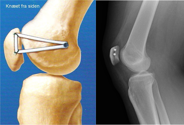
MPFL is a ligament that attaches the kneecap to the femoral condyle on the inner side of thigh and is responsible for stabilising the patella to glide on the trochlea especially in the initial stages of knee bending. The function of this ligament can be compared to the reins of a horse – Just like the reins of a horse control the movement of the horse, the MPFL controls the movement of the patella in the trochlea. MPFL reconstruction surgery replaces the native MPFL ligament by a stronger ligament that keeps the patella (kneecap) in place (see image below) . The ligament is typically harvested from the hamstrings (tendons running on the back of the thigh, by an incision over the inner site of the shin bone (tibia).

Tibia Tubercle Osteotomy (TTO)
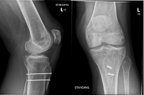
This procedure involves the surgical movement of the tibial tuberosity (the area on the shin bone where the patellar tendon attaches) either medially (inwards) or inferiorly or both depending on the risk factors present. The fixation is held with 2 or 3 screws to hold the repositioned new bone in its new position. It takes 3-4 months for the bones to heal completely

How is the diagnosis made?
Diagnosis is often made by an MRI scan which reveals the integrity of the MPFL ligament. It is also useful to diagnose risk factors including high riding patella (patella alta) and increased TT-TG distance (tibial tuberosity – trochlear groove), the two parameters which are often associated with patellar instability.
How is the surgery done?
The procedure is usually done under general anesthesia or regional anesthesia. The procedure usually takes between 60-90 minutes and done under a tourniquet. The hamstring tendon is harvested first. The central part of the tendon is then buried in a grove on the inner side of the patella while the tails are brought out and inserted into the inner side of the femur at a fixed point identified with the help of x-ray, these tails are then inserted into the drill hole on the femur and held in place with an absorbable screw.
Rehabilitation
This is usually done as a day case procedure. You should be able to go home the same day. Weight bearing status depends on whether you have had TTO procedure along with MPFL. If only MPFL surgery was done, you can fully weight bear immediately after the surgery and can bend the knee earlier. However, if a TTO surgery is done, then you will have a brace that will control the amount of knee bend in the first 6 weeks to protect the osteotomy site. Further you will non weight bearing for the first 6 weeks and weight bearing status is determined by the degree of healing of the bone. You will receive physiotherapy to strengthen the muscles and improve the range of movement of the knee.
Return to Normality
You should be able to return to desk job at 6-8 weeks while normal activities can be completely resumed usually at 3-4 months. Driving is usually possible at 6-8 weeks. Unrestricted sporting activities are usually resumed at 5-6 months.
Complications
Like any other surgery, this procedure also has its own complications. In addition to the routine complications with any surgical procedure like infection and clot in legs (DVT)/lung clot (PE), complications specific to this procedure are recurrence of dislocation (between 3-5%), screw breakage and need to replace them, knee stiffness (1-3%), failure of bone to heal (more common in smokers) and necessity for revision surgery in a very small number of patients.
